


Clarence Collison
Honey Bee Digestive/
Excretory System Functions
By: Clarence Collison
“The worker honey bee ingests 4 substances: nectar, honeydew, pollen and water (Morse and Hooper, 1985b).” “The digestive system of the bee has two main functions, to provide for the nutritional needs of the bee itself and also as a means of storage and temporary transport of nectar back to the hive. Nectar, which is to be returned to the hive, is stored in the crop and is prevented from passing further through the digestive system. However, pollen contained within the nectar can be strained out and selectively digested for the bees’ own needs (Stell 2012).”
“The function of the excretory system is to maintain a constant internal environment in the body, by the elimination or segregation of unwanted substances present in the blood, and by the retention or reabsorption of constituents needful to the organism. Many organs and tissues contribute to this end, but the most important are the Malpighian tubes, which discharge into the intestine at the junction of the mid-gut with the hind-gut. The urine, the product of the Malpighian tubes, is commonly mixed with the residue of food from the mid-gut; these together make up the excrement or feces of insects (Wigglesworth, 1972).”
“The main components of the digestive system begin with the salivary glands in both the head and thorax, which add enzymes to food in the mouth. This mixture is then carried up through the pharynx before passing below the brain, through the neck and along the esophagus in the lower part of the thorax. The esophagus then enters the abdomen through the petiole just above the ventral diaphragm. In the abdomen the esophagus widens out into the crop (honey stomach), which occupies when full, much of the front and lower part of the abdomen. The main digestive organ is the ventriculus. This draws food out of the crop by means of a mouth-like structure, the proventriculus. Food in the ventriculus is digested by enzymes here before passing on to the small intestine (Stell, 2012).”
“The honey bee intestine is distinctly divided into two parts: first a narrow anterior intestine which is looped or coiled in various ways according to the position of the stomach; and second, a large pear-shaped posterior intestine or rectum, opening by its tapering extremity through the anus into the cavity that contains the sting. The intestine and rectum serve principally for the discharge of waste matter and for the absorption of water, but the rectum is also a storage chamber for the retention of feces until the latter can be evacuated outside the hive. In overwintering bees, the rectum may become so greatly distended as to occupy a large part of the abdominal cavity before defecation occurs (Snodgrass and Erickson, 1992).”
“At the junction of the intestine with the ventriculus, a great number of long, thread-like tubes, probably a hundred or more of them, open into the intestine. These tubes are known as the Malpighian tubules; they are not digestive glands but excretory organs that remove waste products of metabolism from the hemolymph, including both nitrogenous substances and salts (Snodgrass and Erickson, 1992).” “They are also osmoregulatory organs; maintaining water and ion balance in the medium surrounding vital cells and organs (Lipiński, 2018).” “The tubules extend long distances in the body cavity, winding and twisting in numerous convolutions through the spaces about the other organs, where they are directly bathed by the blood. The products discharged into the intestine are eliminated along with the waste food matter (Snodgrass and Erickson, 1992).”
“The alimentary canal carries out many functions critical to insect physiology, including digesting and absorbing nutrients and water, housing beneficial gut bacteria, and eliciting immunological responses. The midgut, in particular, is a compartment for digestion and absorption and serves as a first point of contact between ingested pathogens and the insect immune system. Recently, the protein vitellogenin (Vg) was localized in midgut cells of some honey bee workers. Vg is an important egg-yolk precursor protein in nearly all oviparous animals, but it also has immunological functions shared across many taxa. Additional and unexpected Vg functions have been characterized in honey bees, but none of these functions involve the midgut directly. Therefore, we sought to map out how Vg is localized and expressed in this organ across the two most common worker bee behavioral groups, namely nurses and foragers. We used immunohistochemistry and quantitative reverse transcription PCR to show Vg has different localization patterns in nurses and foragers and limited gene expression. Our study provides a platform for building a more detailed understanding of the possible roles of Vg in insect midgut cells, and it adds to the current knowledge of this fascinating, multi-functional protein (Harwood and Amdam, 2021).”
“The food within the ventriculus is contained within thin-walled membranous bags known as peritrophic membranes. This membrane is secreted by cells at the beginning of the ventriculus when food first enters. The digestive enzymes are able to pass through the membrane to reach the food and the nutrients are able to pass out. It is thought that this membrane provides protection against physical abrasion to the lining of the ventriculus from rough-edged particles, restricts bacteria and other potentially harmful small organisms and concentrates the digestive process (Stell, 2012).”
“For storage of food reserves, bees have cream-colored cells on the dorsal and ventral parts of the abdomen called fat bodies. These cells concentrate and store fat, protein in the form of albumen, and glycogen, which can be converted to glucose when needed (Winston, 1987).”
“All undigested material passes from the small intestine into the rectum to be stored prior to defecation. “The undigested solid wastes, consists primarily of pollen husks, fat globules, and dead midgut cells (Winston, 1987).” “Bees never eject their feces in the hive (Snodgrass, 1956).”
“In the anterior part of the rectum are six long, regular spaced thickenings of the epithelial wall, which are characteristic features of the rectum in most adult insects and formerly were known as “rectal glands.” Since no secretory activity has ever been observed in these organs, they are now termed rectal pads. When the rectum of the bee is distended, the pads appear on the outer surface as six opaque ridges (Snodgrass, 1956).” “The rectal pads are responsible for absorption of water and salts (Santos et al., 2009).”
The digestive/excretory systems of the larval honey bee are much simpler than that of adult workers. “The main purpose of the honey bee larval stage is to eat and grow, and therefore internally the alimentary canal takes up most of the space. The alimentary canal comprises the stomodeum, the ventriculus, where digestion takes place, and the proctodeum. The proctodeum, or hind gut, is not connected to the ventriculus until just before the larva pupates. This adaptation is because the larvae tend to lie on and amongst their food, which thus does not become fouled with excretory products until they have finished feeding. The four large Malpighian tubules remove waste products from the blood — mainly excess nitrogen from the breakdown of proteins — and store it, mainly as uric acid, until they discharge into the ventriculus when this breaks through into the hindgut, and from there to the outside, just before pupation (Morse and Hooper, 1985a).”
“The last few days of larval life are spent constructing a cocoon within the cell. To spin the cocoon, the larvae uncurl and stretch out fully in the cells with their heads towards the capped end (Jay, 1963) and begin weaving the cocoon with their spinnerets. The main substance used in the cocoon is silk, secreted from what will become, the thoracic salivary glands in the adults. The larvae also defecate early in cocoon construction; the excretory tubules and midgut are closed off until feeding is completed, when their contents are just discharged into the base of the cell. The dark brown feces as well as a lighter colored substance from the excretory tubules make up most of the other materials used in cocoon construction (Jay, 1964) (Winston, 1987).”
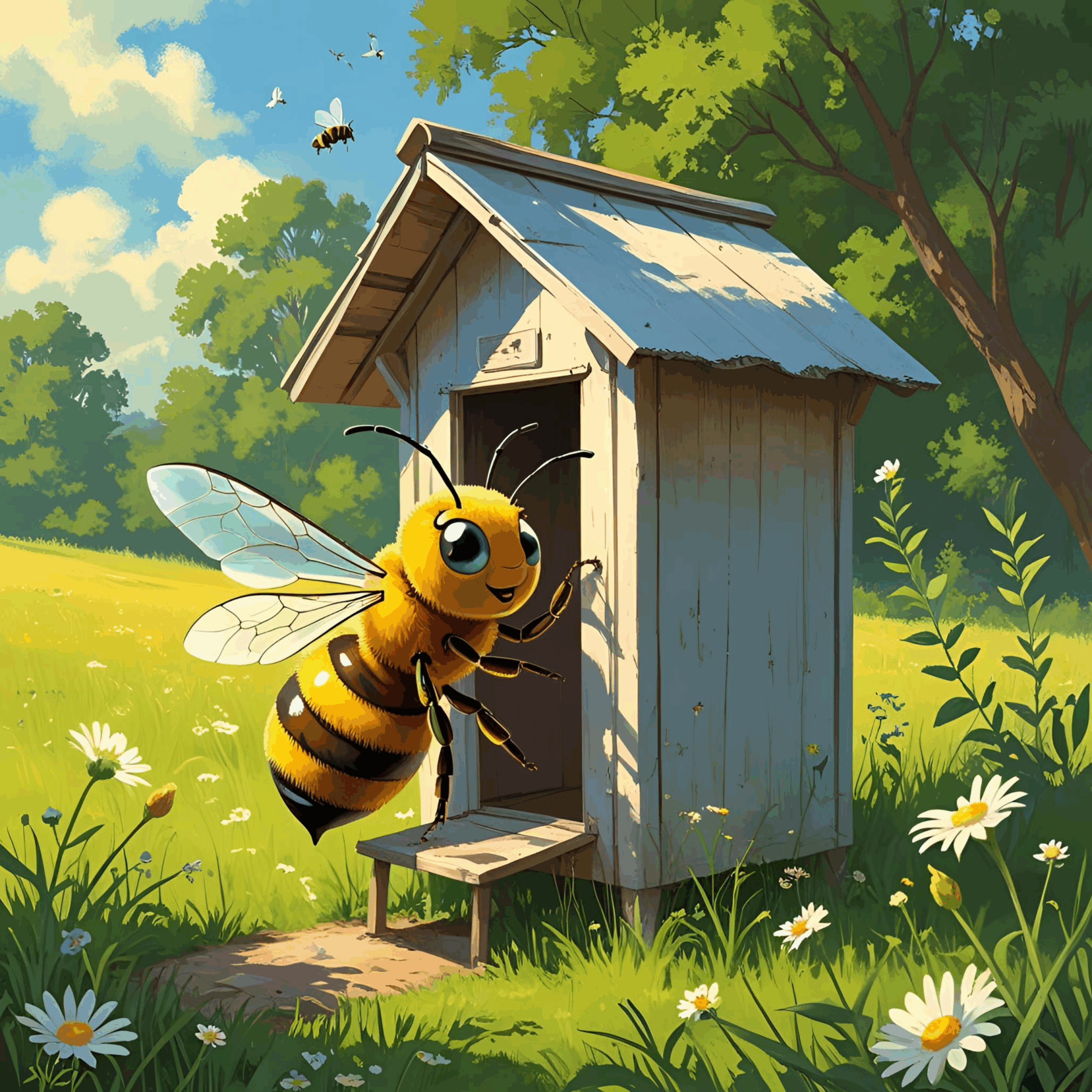
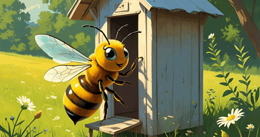
“A honey bee using an outhouse,” image generated by Canva’s Magic Media Tool, August 5, 2025. © Canva.com.
“Studies of newly emerged honey bee workers have demonstrated that their guts are colonized by a consistent core microbiota within several days of eclosure. Experiments were conducted aimed at illuminating the transmission routes and spatiotemporal colonization dynamics of this microbiota. Experimental groups of newly emerged workers were maintained in cup cages and exposed to different potential transmission sources. Colonization patterns were evaluated using quantitative real-time PCR (qPCR) to assess community sizes and using deep sequencing of 16S rRNA gene amplicons to assess community composition. In addition, they monitored the establishment of the ileum and rectum communities within workers sampled overtime from natural hive conditions. The study verified that workers initially lack gut bacteria and gain large characteristic communities in the ileum and rectum within 4 to 6 days within hives. Typical communities, resembling those of workers within hives, were established in the presence of nurse workers or nurse worker fecal material, and atypical communities of noncore or highly skewed compositions were established when workers were exposed only to oral trophallaxis or hive components (comb, honey, bee bread). The core species of Gram-negative bacteria, Snodgrassella alvi, Gilliamella apicola, and Frischella perrara, were dependent on the presence of nurses or hindgut material, whereas some Gram-positive species were more often transferred through exposure to hive components. These results indicate aspects of the colony life cycle and behavior that are key to the propagation of the characteristic honey bee gut microbiota (Powell et al., 2014).”
“Newly emerged honey bees were fed diets containing 5% protein in the form of dandelion pollen, egg albumin, soybean meal hydrolysate or casein for 7 days, after which the recta were removed and the nitrogenous excretory products determined in the fecal material. Bees fed pollen showed the following as percentage of total nitrogen: uric acid, 62.06; creatine, 1.5; creatinine, 1.47; amino nitrogen, 6.11; urea, 0; and ammonia, 0. Amino nitrogen showed a direct relationship to diet adequacy: 1.36 and 6.11% for the negative controls and pollen-fed groups, respectively. No consistent pattern was noted for creatine or creatinine, except that both were lower for the pollen-fed groups than for the negative controls. When dietary protein sources were arranged in order of decreasing adequacy based on pharyngeal gland development, uric acid excretion varied inversely with diet adequacy; pollen, 48 (percentage of total nitrogen excreted); egg albumin, 62; soy hydrolysate, 62; casein, 96; and negative control, 96 (McNally et al., 1965).”
“Malpighamoeba mellificae is a protozoan that infects the Malpighian tubules of honey bees. The amoebae, ingested as cysts, develop into trophozoites that feed upon tubule epithelia. The resulting damage of the Malpighian tubules can induce an imbalance of waste excretion and hemolymph exchange. This causes the so-called amoebiasis disease in adult bees, which may co-occur with Nosema infections. Most reports of this amoeba are from the 1960s and earlier, and knowledge of the disease and its spreading is very poor. The lack of any genetic marker for the species hampers its sensitive identification using molecular tools and gaining knowledge on its epidemiology. Here, we present a diagnostic RT-qPCR assay, consisting of two primers and one probe that were developed based on 18S rRNA sequences of the amoeba, generated with metagenomic sequencing of Malpighian tubules with and without M. mellificae cysts. The assay was initially tested and adjusted with samples microscopically tested for the presence of M. mellificae cysts. Later, it was validated and material with unknown infection status was tested. The sensitive diagnostic Malpighamoeba disease 18S assay is now ready to be applied for honey bee health monitoring purposes and to investigate the prevalence of M. mellificae in more detail (Schäfer et al., 2022).”
“Knowledge of the spreading mechanism of honey bee pathogens within the hive is crucial to our understanding of bee disease dynamics. The aim of this study was to assess the presence of infectious chronic bee paralysis virus (CBPV) in bee excreta and evaluate its possible role as an indirect route of infection. Samples of paralyzed bees were (i) produced by experimental inoculation with purified virus and (ii) collected from hives exhibiting chronic paralysis. CBPV in bee heads or feces (crude or absorbed onto paper) was detected by reverse transcription-PCR. CBPV infectivity was assessed by intrathoracic inoculation of bees with virus extracted from feces and by placement of naive bees in cages previously occupied by contaminated individuals. CBPV RNA was systematically detected in the feces of naturally and experimentally infected bees and on the paper sheets that had been used to cover the floors of units containing bees artificially infected with CBPV or the floor of one naturally infected colony. Both intrathoracic inoculation of bees with virus extracted from feces and placement of bees in contaminated cages provoked overt disease in naive bees, thereby proving that the excreted virus was infectious and that this indirect route of infection could lead to overt chronic paralysis. This is the first experimental confirmation that infectious CBPV particles excreted in the feces of infected bees can infect naive bees and provoke overt disease by mere confinement of naive bees in a soiled environment (Ribière et al., 2007).”
References
Harwood, G. and G. Amdam 2021. Vitellogenin in the honey bee midgut. Apidologie 52: 837-847.
Jay, S.C. 1963. The longitudinal orientation of the larval honeybees (Apis mellifera) in their cells. Can. J. Zool. 41: 717-723.
Jay, S.C. 1964. The cocoon of the honeybee, Apis mellifera L, Can. Entomol. 96: 784-792.
Lipiński, Z. 2018. Honey Bee Nutrition And Feeding. OZGraf S.A., Olsztyn, Poland, 430 pp.
McNally, J.B., W.F. McCaughey, L. N. Standifer and F.E. Todd 1965. Partition of excreted nitrogen from honey bees fed various proteins. J. Nutr. 85: 113-116.
Morse, R.A. and T. Hooper 1985a. Larva, In The Illustrated Encyclopedia Of Beekeeping. pp. 233-235. Edited by R. Morse and T. Hooper. E. P. Dutton, Inc. New York, NY.
Morse, R.A. and T. Hooper 1985b. Nutrition of the honey bee, In The Illustrated Encyclopedia Of Beekeeping. pp. 280-281. Edited by R. Morse and T. Hooper. E. P. Dutton, Inc. New York, NY.
Powell, J.E., V.G. Martinson, K. Urban-Mead and N.A. Moran 2014. Routes of acquisition of the gut microbiota of the honey bee Apis mellifera. Appl. Environ. Microbiol. 80: 7378-7387.
Ribière, M., P. Lallemand, A.-L. Iscache, F. Schurr, O. Celle, P. Blanchard, V. Olivier and J.-P. Faucon 2007. Spread of infectious chronic bee paralysis virus by honeybee (Apis mellifera L.) feces. Appl. Environ. Microbiol. 73: 7711-7716.
Santos, C.G., C.A. Neves, J.C. Zanuncio and J.E. Serrāo 2009. Postembryonic development of rectal pads in bees (Hymenoptera: Apidae). Anat. Rec. 292: 1602-1611.
Schäfer, M.O., J. Horenk and C. Wylezich 2022. Molecular detection of Malpighamoeba mellificae in honey bees. Vet. Sci. 9: 148. https:// doi.org/10.3390/vetsci9030148
Snodgrass, R.E. 1956. Anatomy Of The Honey Bee, Comstock Publ. Assoc., Ithaca, NY, 2nd ed.
Snodgrass, R.E. and E.H. Erickson 1992. The anatomy of the honey bee. In The Hive And The Honey Bee, Dadant and Sons, Inc., Hamilton, IL. pp. 103-169.
Stell, I. 2012. Understanding Bee Anatomy: A Full Colour Guide. The Catford Press, Teddington, Middlesex, UK, 203 pp.
Wigglesworth, V.B. 1972. Excretion. In: The Principles of Insect Physiology. Springer, Dordrecht. pp. 553-592. https://doi.org/10.1007/978-94-009-5973-6_12
Winston, M.L. 1987. The Biology Of The Honey Bee. Harvard University Press, Cambridge, MA, 281 pp.
Clarence Collison is an Emeritus Professor of Entomology and Department Head Emeritus of Entomology and Plant Pathology at Mississippi State University, Mississippi State, MS. Became interested in entomology through 4-H and studied honey bees for both my Master’s and Ph.D. Have been involved with beekeeping for 50+ years.