Immunity Mechanisms and Immunosenescence
By: Clarence Collison
 “Honey bees face many important parasites and pathogens against which they have evolved behavioral, morphological, physiological and immune based defenses” (Evans, 2006). At the individual level, immune mechanisms are comprised of: 1) resistance mechanisms associated with anatomical and physiological barriers of the body, 2) cell-mediated immunity involving hemocytes (blood cells, including plasmocytes, lamellocytes and granulocytes), 3a) congenital humoral resistance related to the activity of lysozyme (N-acetylmuramylhydrolase), the prophenoloxidase system and hemagglutinins (lectins), and 3b) induced humoral resistance based on the action of antimicrobial peptides: abaecin, apidaecin, hymenoptaecin and defensin. In addition to the individual resistance of each bee, there are also defense mechanisms activated at the colony level. Shared secretion resistance is connected with the presence of antipathogenic compounds in secreta and in bee products, i.e. propolis. Social immunity is associated with hygienic and nursing behaviors, as well as with age polyethism in the colony, swarming and the changing behavior of sick individuals (Strachecka et al., 2018).
The innate immune system includes the circulating hemocytes (immune cells) that clear pathogens from hemolymph (blood) by phagocytosis, nodulation or encapsulation. Honey bee hemocyte numbers have been linked to hemolymph levels of vitellogenin. Vitellogenin is a multifunctional protein with immune-supportive functions identified in a range of species, including the honey bee. Hemocyte numbers can increase via mitosis (cell division), and this recruitment process can be important for immune system function and maintenance. Hystad et al. (2017) tested to see if hemocyte mediated phagocytosis (engulfing of microorganisms) differs among the physiologically different honey bee worker castes (nurses, foragers and Winter bees), and studied possible interactions with vitellogenin and hemocyte recruitment. They found that nurses are more efficient in phagocytic uptake than both foragers and Winter bees. Vitellogenin was detected within the hemocytes and they found that Winter bees have the highest numbers of vitellogenin positive hemocytes. Connections between phagocytosis, hemocyte-vitellogenin and mitosis (cell division) were worker caste dependent. Their results demonstrate that the phagocytic performance of immune cells differs significantly between honey bee worker castes, and support increased immune competence in nurses as compared to forager bees. Their data also provides support for roles of vitellogenin in hemocyte activity.
As honey bees mature, the types of pathogens they experience also change. As such, pathogen pressure may affect bees differently throughout their lifespan. Wilson-Rich et al. (2008) investigated immune strength across four developmental stages: larvae, pupae, nurses (one day old adults) and foragers (22-30 day old adults). The immune strength of honey bees was quantified using standard immunocompetence assays: total hemocyte count, encapsulation response, fat body quantification and phenoloxidase activity. Larvae and pupae had the highest total hemocyte counts, while there was no difference in encapsulation response between developmental stages. Nurses had more fat body mass than foragers, while phenoloxidase activity increased directly with honey bee development. Immune strength was most vigorous in older, foraging bees and weakest in young bees. Importantly, they found that adult honey bees do not abandon cellular immunocompetence as was recently proposed. Induced shifts in behavioral roles may increase a colony’s susceptibility to disease if nurses begin foraging activity prematurely.
Male and female bees are subject to differing selective pressures due to their differences in colony tasks and changes in the threat of pathogen infection at different life stages. Laughton et al. (2011) investigated the immune response of workers and drones at all developmental phases, from larvae through to late stage adults, assaying both a constitutive (phenoloxidase, PO activity) and induced (antimicrobial peptide, AMP) immune response. They found that larval bees have low levels of PO activity. Adult workers produced stronger immune responses than drones, and a greater plasticity in immune investment. An immune challenge resulted in lower levels of PO activity in adult workers, which may be due to the rapid utilization and a subsequent failure to replenish the constitutive phenoloxidase. Both adult workers and drones responded to an immune challenge by producing higher titers of AMPs, suggesting that the cost of this response prohibits its constant maintenance. Both castes showed signs of senescence in immune investment in the AMP response. Different sexes and life stages therefore alter their immune system management based on the combined factors of disease risk and life history.
Randolt et al. (2008) employed the proteomic approach in combination with mass spectrometry to study the immune response of honey bee workers at different developmental stages. Analysis of the hemolymph proteins of non-infected, mock-infected and immune-challenged individuals by polyacrylamide gel electrophoresis showed differences in the protein profiles. They present evidence that in vitro reared honey bee larvae respond with a prominent humoral reaction to aseptic and septic injury as documented by the transient synthesis of the three antimicrobial peptides (AMPs) hymenoptaecin, defensin 1 and abaecin. In contrast, young adult workers react with a broader spectrum of immune reactions that include the activation of prophenoloxidase and humoral immune responses. At least seven proteins appeared consistently in the hemolymph of immune-challenged bees, three of which are identical to the AMPs induced also in larvae. The other four, i.e., phenoloxidase (PO), peptidoglycan recognition protein-S2, carboxylesterase (CE) and an Apis-specific protein not assigned to any function (HP30), are induced specifically in adult bees and, with the exception of PO, are not expressed after aseptic injury. Structural features of CE and HP30, such as classical leucine zipper motifs, together with their strong simultaneous induction upon challenge with bacteria suggest an important role of the two novel bee-specific immune proteins in response to microbial infections.
Female insects that survive a pathogen attack can produce more pathogen-resistant offspring in a process called trans-generational immune priming. In the honey bee, the egg-yolk precursor protein vitellogenin transports fragments of pathogen cells into the egg, thereby setting the stage for a recruitment of immunological defenses prior to hatching. Honey bees live in complex societies where reproduction and communal tasks are divided between a queen and her sterile female workers. Worker bees metabolize vitellogenin to synthesize royal jelly, a protein-rich glandular secretion fed to the queen and young larvae. Harwood et al. (2019) investigated if workers can participate in trans-generational immune priming by transferring pathogen fragments to the queen or larvae via royal jelly. As a first step toward answering this question, they tested whether worker-ingested bacterial fragments can be transported to jelly-producing glands, and what role vitellogenin plays in this transport. To do this, they fed fluorescently labeled Escherichia coli to workers with experimentally manipulated levels of vitellogenin. They found that bacterial fragments were transported to the glands of control workers, while they were not detected at the glands of workers subjected to RNA interference-mediated vitellogenin gene knockdown, suggesting that vitellogenin plays a role in this transport. Their results provide initial evidence that trans-generational immune priming may operate at a colony-wide level in honey bees.
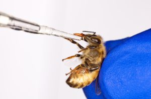
Honey bee larvae are highly susceptible to the bacterial pathogen Paenibacillus larvae (causative agent of American foulbrood) only during the first instar of larval development. Transcript levels were measured for genes encoding two antimicrobial peptides, abaecin and defensin, as well as two candidates in the immune response cascade (PGRP-LD and masquerade) in control larvae and larvae exposed to the pathogen. Transcripts (a length of RNA or DNA that has been transcribed respectively from a RNA or DNA template) for all four are present throughout development. This suggests that other physiological or dietary factors may better explain the age-based change in vulnerability to this pathogen. One of these genes, abaecin, shows significant up-regulation 24 hours following oral inoculation with P. larvae, precisely when the bacterium surmounts the midgut epithelia of bees. Expression of both antimicrobial peptides varied by 1000-fold across different nestmate bees, indicating an allelic component to their expression (Evans, 2004).
An example of immunosenescence is seen in the worker caste. The bee’s age-associated transition from hive duties to more risky foraging activities is linked to a dramatic decline in immunity. Explicitly, it has been shown that an increase in the juvenile hormone (JH) level, which accompanies onset of foraging behavior, induces extensive hemocyte death through nuclear pycnosis (degeneration of cell nucleus). Amdam et al. (2005) demonstrated that foragers that are forced to revert to hive-tasks showed reversal of immunosenescence, i.e. a recovery of immunity with age. This recovery, which is triggered by a social manipulation, is accompanied by a drop in the endogenous JH titer and an increase in the hemolymph vitellogenin level.
They also established that worker immunosenescence is mediated by apoptosis (the death of cells), corroborating that reversal of immunosenescence emerges through proliferation of new cells. The results reveal a unique flexibility in honey bee immunity.
Immunosenescence, the systemic reduction of immune efficiency with age, is increasingly recognized as having important implications for host-parasite dynamics. Changes in the immune response can impact the ability of an individual to resist or moderate parasite infection, depending on how and when it encounters a parasite challenge. Using the European honey bee Apis mellifera and its microsporidian parasite Nosema ceranae, we investigated the effects of host age on the ability to resist parasite infection and on baseline immunocompetence, assessed by quantifying constitutive (PO) and potential levels (PPO) of the phenoloxidase immune enzyme as general measures of immune function. There was a significant correlation between the level of general immune function and infection intensity, but not with survival, and changes in immune function with age correlated with the ability of individuals to resist parasite infection. Older individuals had better survival when challenged with a parasite than younger individuals, however they also had more intense infections and lower baseline immunocomptence. The ability of older individuals to have high infection intensities yet live longer, has potential consequences for parasite transmission (Roberts and Hughes, 2014).
Young honey bee workers (zero to two to three weeks old) perform tasks inside the colony, including brood care (nursing), whereas older workers undergo foraging tasks during the next three to four weeks, when an intrinsic senescence program culminates in worker death. It was hypothesized that foragers are less able to react to immune system stimulation than nurse bees and that this difference is due to an inefficient immune response in foragers. To test this hypothesis, an experimental design was used that allowed them to uncouple chronological age and behavior status (nursing/foraging). Worker bees from a normal age demography colony (where workers naturally transit from nursing to foraging tasks as they age) and of a single-cohort colony setup (composed of same-aged workers performing nursing or foraging tasks) were tested for survival and capability of activation of the immune system after bacterial injection. Expression of an antimicrobial peptide gene, defensin-1 (def-1), was used to assess immune system activation. They then checked whether the immune response includes changes in the expression of aging- and behavior-related genes, specifically vitellogenin (vg), juvenile hormone esterase (jhe), and insulin-like peptide-1 (ilp-1). A significant difference was found in survival rate between bees of different ages but carrying out the same tasks. The results thus indicate that the bees’ immune response is negatively affected by intrinsic senescence. Additionally, independent of age, foragers had a shorter lifespan than nurses after bacterial infection, although both were able to induce def-1 transcription. In the normal age demography colony, the immune system activation resulted in a reduction in the expression of vg, jhe and ilp-1 genes in foragers, but not in the nurse bees, demonstrating that age and behavior are both important influences on the bees’ immune response. By disentangling the effects of age and behavior in the single-cohort colony, it was found that vg, jhe and ilp-1 response to immune system stimulation was independent of behavior. Younger bees were able to mount a stronger immune response than older bees, thus highlighting age as an important factor for immunity. Taken together, the results provide new insights into how age and behavior affect the honey bee’s immune response (Lourenco et al., 2019).
Foragers facilitate horizontal pathogen transmission in honey bee colonies, yet their systemic immune function wanes during transition to this life stage. In general, the insect immune system can be categorized into mechanisms operating at both the barrier epithelial surfaces and at the systemic level. As proposed by the intergenerational transfer theory of aging, such immunosenescence may result from changes in group resource allocation. Yet, the relative influence of pathogen transmission and resource allocation on immune function in bees from different stages has not been examined in the context of barrier immunity. They found that expression levels of antimicrobial peptides (AMPs) in honey bee barrier epithelia of the digestive tract do not follow a life-stage-dependent decrease. In addition, correlation of AMP transcript abundance with microbe levels reveals a number of microbe-associated changes in AMPs levels that are equivalent between nurses and foragers. These results favor a model in which barrier effectors are maintained in foragers as a first line of defense, while systemic immune effectors are dismantled to optimize hive-level resources (Jefferson et al., 2013).
References
Amdam, G.V., A. Aase, S.C. Seehuus, M.K. Fondrk and K. Hartfelder 2005. Social reversal of immunosenescence in honey bee workers. Exp. Gerontol. 40: 939-947.
Evans, J.D. 2004. Transcriptional immune responses by honey bee larvae during invasion by the bacterial pathogen, Paenibacillus larvae. J. Invertebr. Pathol. 85: 105-111.
Evans, J.D. 2006. Beepath: an ordered quantitative-PCR array for exploring honey bee immunity and disease. J. Invertebr. Pathol. 93: 135-139.
Harwood, G., G. Amdam and D. Freitak 2019. The role of vitellogenin in the transfer of immune elicitors from gut to hypopharyngeal glands in honey bees (Apis mellifera). J. Insect Physiol. 112: 90-100.
Hystad, E.M., H. Salmela, G.V. Amdam and D. Münch 2017. Hemocyte-mediated phagocytosis differs between honey bee (Apis mellifera) worker castes. PLoS ONE 12(9): e0184108.
Jefferson, J.M., H.A. Dolstad, M.D. Sivalingam and J.W. Snow 2013. Barrier immune effectors are maintained during transition from nurse to forager in the honey bee. PLoS ONE 8(1): e54097.
Laughton, A.M., M. Boots, and M.T. Silva-Jothy 2011. The ontogeny of immunity in the honey bee, Apis mellifera L. following an immune challenge. J. Insect Physiol. 57: 1023-1032.
Lourenco, A.P., J.R. Martins, F.A.S. Torres, A. Mackert, L.R. Aguiar, K. Hartfelder, M.M.G. Bitondi and Z.L.P. Simões 2019. Immunosenescence in honey bees (Apis mellifera L.) is caused by intrinsic senescence and behavioral physiology. Exp. Gerontol. 119: 174-183.
Randolt, K., O. Gimple, J. Geissendörfer, J. Reinders, C. Prusko, M.J. Mueller, S. Albert, J. Tautz and H. Beier 2008. Immune-related proteins induced in the hemolymph after aseptic and septic injury differ in honey bee worker larvae and adults. Arch. Insect Biochem. Physiol. 69: 155-167.
Roberts, K.E. and W.O.H. Hughes 2014. Immunosenescence and resistance to parasite infection in the honey bee, Apis mellifera. J. Invertebr. Pathol. 121: 1-6.
Strachecka, A., A. Los, J. Filipczuk, and M. Schulz 2018. Individual and social immune mechanisms of the honey bee. Med. Weter. 74: 426-433.
Wilson-Rich, N., S.T. Dres and P.T. Starks 2008. The ontogeny of immunity: development of innate immune strength in the honey bee (Apis mellifera). J. Insect Physiol. 54: 1392-1399.
Clarence Collison is an Emeritus Professor of Entomology and Department Head Emeritus of Entomology and Plant Pathology at Mississippi State University, Mississippi State, MS.


