


Click Here if you listened. We’d love to know what you think. There is even a spot for feedback!
Read along below!

Found in Translation
DNA Fingerprinting
By: Jay Evans, USDA Beltsville Bee Lab
Long before your personal fingerprints became a tool to open computers, they were used to help solve crimes and other mysteries. Unique to all but identical twins, fingerprints can be irrefutable evidence of past touches. In fact, many of us bought cheap detective kits with a then-mysterious white powder and tried to solve household crimes with this new tool (hint: your sister wasn’t really using your toothbrush). Currently, stopovers by living beings are marked most accurately by traces of the nucleic acid sequences that make up their genomes. The past decade has shown that DNA and RNA are, for lack of a better word, leaky. So much so that if one wants to screen a meadow for an extremely rare bee species, the current method of choice is to collect some of the flowers in that meadow, isolate bee DNA, and use extremely sensitive genetic screens to divvy up the genomes of all past bee visitors. This is not science fiction. Arndt Schmidt and colleagues used DNA signals to survey bee diversity across flower plots (Schmidt, A., Schilbach, L., Schanowski, A. et al. (2025) Flower-derived environmental DNA reveals community diversity, species abundances and ecological interactions in bee pollinators.” Environmental DNA 7, no. 4: e70178. https://doi.org/10.1002/edn3.70178). Yes, there’s even a journal called Environmental DNA. Their most accurate strategy involved cutting flower heads from 10-12 plants species per field, putting flowers from each species into bags with sterile water, shaking, and then purifying DNA from that water rinse. These scientists identified a remarkable 54 bee species across three plots sampled four times each, a bee diversity score equal to teams of expert field bee experts with nets and good ID skills. This technique is clearly up to the task of knowing which bees were doing the work of pollination (yes, honey bees made the list).
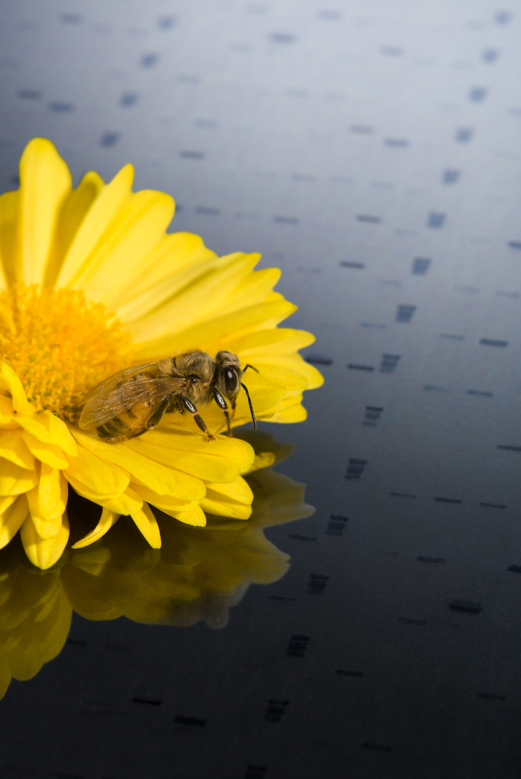
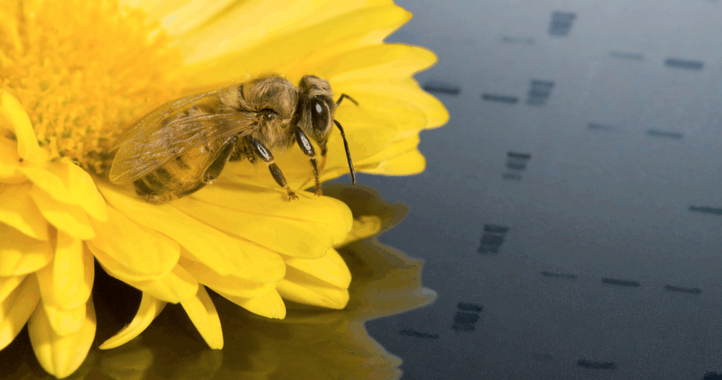
Honey bee on a DNA fragment analysis map. The honey bee genome is being studied to improve reproduction, behavior, disease resistance, and more. Photo by Peggy Greb.
For honey bee health it is most important to identify not the bee herself, but microbes and other associates good or bad carried by that bee. The screening of environmental DNA (eDNA) is more that up for this task. A key question is which hive material is best for screening. Choices range from bee bodies to hive stores, and from dirty bottom boards to surrounding flowers. While leaky DNA signals have been found in all, there are new insights into the best substrate to study. Jessica Henneken and colleagues in Australia pushed the limits of bottom-board debris to characterize bacteria, fungi and arthropods (insects and mites in this case) that had sometime in the past oozed or crawled across the bottoms of bee hives (Henneken, J., F. Martoni, A. Piper, R. Smith, and B. Rodoni. (2025) Molecular surveillance of honey bee hives using eDNA metabarcoding during pollination season.” Environmental DNA 7, no. 6: e70220. https://doi.org/10.1002/edn3.70220). In this study, thousands of hive visitors were identified, including wax moths and hive beetles, while a dozen potential threats (including Tropilaelaps mites) were correctly shown to be absent in sampled DNA. This study builds on earlier bottom-board forensic work by Leigh Boardman and colleagues at the University of Florida (Boardman, L., Marcelino, J. A. P., Valentin, R. E., Boncristiani, H., Standley, J. M., & Ellis, J. D. (2024). Novel eDNA approaches to monitor Western honey bee (Apis mellifera L.) microbial and arthropod communities. Environmental DNA, 6, e419. https://doi.org/10.1002/edn3.419). Both studies relied on massive libraries of DNA sequences for microbes and other hive inhabitants house at the US National Institutes of Health. As those libraries of sequences improve rapidly the ability to scout out almost any life form will itself improve.
Many of us are focused on life forms that do not carry DNA at all, namely the important RNA viruses that devastate honey bee colonies, like Deformed wing virus and paralytic viruses. RNA is generally less stable than DNA, so our go-to method has been to collect live bees and then deep-freeze them until they are processed. Still, since we know viruses can be infective from hive tools and hive stores, their RNA can’t be all that fragile. Indeed, Delka Salkova and colleagues used an RNA capture technique to discover bee viruses (Salkova, D. Balkanska, R. Shumkova, R. Lazarova, S. Radoslavov, G. Hristov, P. (2024) Molecular detection and phylogenetic relationships of honey bee-associated viruses in bee products. Vet. Sci. 11, 369. https:// doi.org/10.3390/vetsci11080369). They used pollen, bee bread, and royal jelly as sources to detect viruses, with slightly more success using the royal jelly component. They obtained valid RNA sequences for the range of bee viruses but were not able to get accurate data for virus levels in colonies. If the intent is both to identify viruses and determine when these viruses are causing colony harm, what is the best way to do that without sampling bees themselves? Fortunately, Erin Hill and colleagues carried out controlled experiments to test just that question (Hill E., Milla L., Encinas-Viso F., O’Dwyer J., Gooden B., Hopper M., Roberts J. (2025) Surface swabs outperform most traditional honey bee (Apis mellifera) hive samples for recovery of eDNA and eRNA. Metabarcoding and Metagenomics 9: e165436. https://doi.org/10.3897/mbmg.9.165436). They found that swabs at the hive entrance adequately capture viruses of bees (along with viruses infecting other organisms large and small), and this was for sure the easiest collection for beekeepers and others. I am not yet convinced this technique is ready to become the standard for quantifying bee viruses. Aside from the whims of bee behavior (presumably incontinent bees will leave a stronger viral sign), the swabs at hive entrances were rich in plant viruses and other incidentals. Consequently, known insect and mite viruses made up maybe one twelfth of the virus pool, dwarfed by viruses carried by plants and fungi. Nevertheless, even samples of pooled bees showed numerous plant virus hitchhikers, and the authors make a good case that the extra effort to dig deeper when sequencing these samples, in order to get a bead on the bee viruses, is worth it given the ease of collection. Studies like this one and the Boardman/University of Florida group show the steps needed to improve detective work and are well on their way towards a major change in beehive sampling. Even today, such hive swabs are likely precise enough to detect arrivals of new mites like Tropilaelaps, as noted by the authors. Future bee detective work seems like it will converge on the talcum powder dusting ‘studies’ of our youth, giving exciting insights from merely swabbing the hive surface and putting those swabs into better and better machines and databases. This is good news for detecting the biological threats faced by our wonderful bees. It has also opened wholly new insights into the range of organisms, intended or ephemeral and from viruses to beekeepers, that pass through the doors of bee boxes.